
Magnetic Resonance Imaging (MRI) is a powerful medical imaging technique used to diagnose and monitor various health conditions. It produces detailed images of soft tissues, organs, bones, and other internal structures without using radiation. This guide outlines exactly what patients can expect before, during, and after an MRI scan, helping reduce uncertainty and improve preparedness.
What Is an MRI Scan?
An MRI scan is a non-invasive diagnostic tool that uses strong magnetic fields and radio waves to create precise images of the body’s internal systems. MRI scanners do not emit ionising radiation like X-rays or CT scans. The process typically takes between 15 and 60 minutes, depending on the body part being examined and the detail required.
When Is an MRI Needed?
MRI scans are requested by healthcare providers to investigate specific symptoms, confirm a diagnosis, or monitor treatment outcomes. Conditions assessed through MRI include:
| Condition Category | Examples |
|---|---|
| Musculoskeletal | Torn ligaments, slipped discs, joint damage |
| Neurological | Brain tumours, multiple sclerosis, stroke damage |
| Abdominal and Pelvic | Liver disease, uterine fibroids, prostate issues |
| Cardiac | Heart valve conditions, congenital heart defects |
| Oncology | Tumour location, size, and metastasis tracking |
Who Needs an MRI Scan?
MRI scans are typically recommended for patients experiencing unexplained pain, neurological symptoms, or known conditions requiring close monitoring. The procedure is safe for adults, children, and the elderly, though some patients may not be suitable candidates if they have metal implants or severe claustrophobia.
Before the MRI: How to Prepare
To prepare for an MRI scan, follow these steps:
- Inform the clinic of implants or metal devices
Notify your provider if you have pacemakers, cochlear implants, surgical clips, or metal fragments. - Avoid wearing metal
Remove jewellery, hearing aids, watches, and clothing with zippers or metallic threads. - Discuss medications and allergies
Some scans involve contrast dye. Inform staff if you have kidney conditions or contrast sensitivity. - Follow fasting guidelines if required
Certain abdominal scans may require you to avoid eating or drinking for 4 to 6 hours. - Wear comfortable clothing
Loose, cotton garments without metal fasteners are ideal. Gowns are provided if necessary.
Features of Pre-MRI Preparation
| Feature | Description |
|---|---|
| Metal screening | Prevents risk of injury or interference with image clarity |
| Patient questionnaire | Confirms medical history and safety clearance |
| Consent forms | Provides legal and clinical authorisation to proceed |
| Clear instructions | Reduces confusion and ensures smoother scanning process |
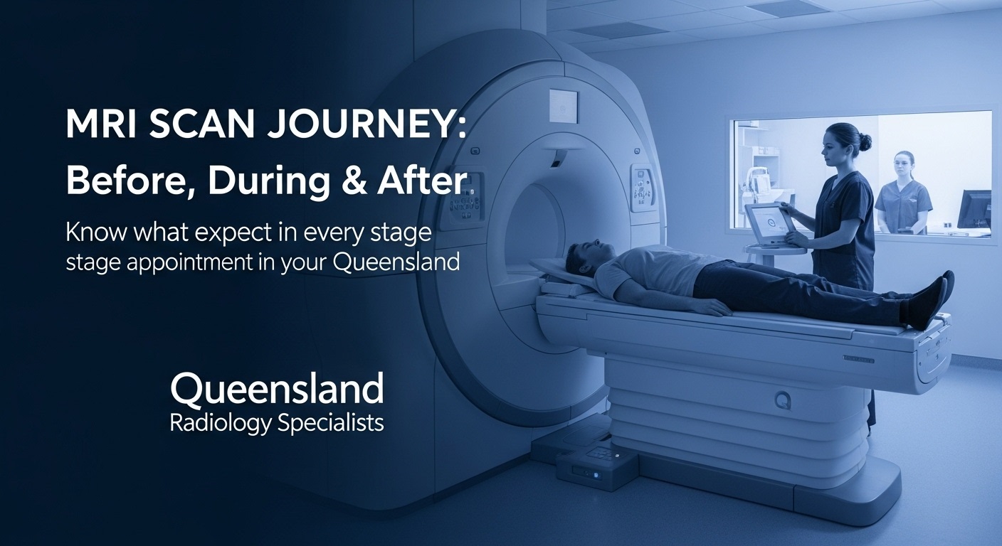
During the MRI: What Happens Inside the Scanner?
To perform an MRI scan, the patient lies flat on a motorised table that slides into a tunnel-like scanner. The machine surrounds the body part being examined.
Here’s what happens during the scan:
- You remain still throughout the scan
Movement can blur images. You may be asked to hold your breath for short periods. - You hear loud tapping or thumping sounds
These are normal noises made by the magnetic coils inside the scanner. - You communicate via intercom
Technicians monitor the process from another room and provide instructions or reassurance. - You may receive a contrast agent
If needed, contrast dye is administered through an intravenous line to improve visibility of blood vessels or soft tissues.
What Does the Scanner Feel Like?
The scanner is not painful. However, the tunnel may feel narrow, especially for those who experience claustrophobia. Some facilities offer open MRI machines for a more spacious experience, or provide mild sedatives upon request.
Functions of the MRI Scanner
| Function | Example Use Case |
|---|---|
| Magnetic field | Aligns hydrogen atoms in the body |
| Radio wave pulses | Disturb the alignment to produce a signal |
| Signal reception | Captures returning signals to build image maps |
| Gradient coils | Help pinpoint image location and orientation |
After the MRI: What Happens Next?
Once your MRI scan is complete, you can resume normal activities unless you received a sedative. There is no recovery period needed for standard scans.
Post-scan steps include:
- Reviewing the images
A radiologist analyses the images and sends a detailed report to your referring doctor. - Collecting results
Most MRI results are available within 24 to 48 hours. Your GP or specialist will discuss the findings. - Following up on treatment
Depending on the scan’s purpose, the results may inform surgical planning, medication adjustments, or further investigations.
Pros and Cons of MRI Scanning
| Pros | Cons |
|---|---|
| Non-invasive and painless | Can feel noisy or claustrophobic for some patients |
| No exposure to radiation | Not suitable for individuals with certain implants |
| Highly detailed images of soft tissue | Takes longer than X-rays or CT scans |
| Widely used for neurological and musculoskeletal issues | Limited access in rural or remote areas |
Situational Relevance: When MRI Is Preferred Over Other Scans
MRI is the preferred imaging choice in situations such as:
- Soft tissue damage (e.g., brain, muscles, tendons)
- Early tumour detection where detailed imaging is critical
- Neurological symptom evaluation such as unexplained seizures or headaches
- Spinal cord injuries and joint pain analysis
In contrast, CT scans are better suited for visualising bone fractures or acute trauma where speed is essential.
Target Audience: Who Should Read This?
This guide is most useful for:
- First-time patients preparing for an MRI
- Caregivers supporting loved ones through medical testing
- GPs referring patients for diagnostic imaging
- Students or healthcare trainees learning about scan protocols
Specific Scenarios: Patient Examples
| Scenario | MRI Use |
|---|---|
| A 55-year-old with lower back pain | MRI detects disc herniation and nerve impingement |
| A 27-year-old athlete with knee pain | MRI shows ACL tear and meniscus damage |
| A 65-year-old post-stroke patient | MRI identifies damage in brain tissue and assists with rehabilitation planning |
MRI at Queensland Radiology Specialists
Queensland Radiology Specialists offer advanced MRI services across multiple locations in Brisbane and regional QLD. Their imaging centres are equipped with high-resolution scanners, experienced radiologists, and patient-focused care protocols. For more information or to book an appointment, visit:
👉 https://www.qldradiologyspecialists.com.au/radiology_services/mri/
Summary: What to Expect During an MRI
To summarise, here’s a step-by-step breakdown:
| Stage | What to Expect |
|---|---|
| Before | Remove metal, complete safety forms, follow instructions |
| During | Lie still, listen to instructions, scanner makes loud noises |
| After | Resume normal activity, results sent to referring doctor within 2 days |
MRI scans are safe, precise, and valuable diagnostic tools. Understanding each phase—before, during, and after—can ease anxiety and support informed medical decisions.